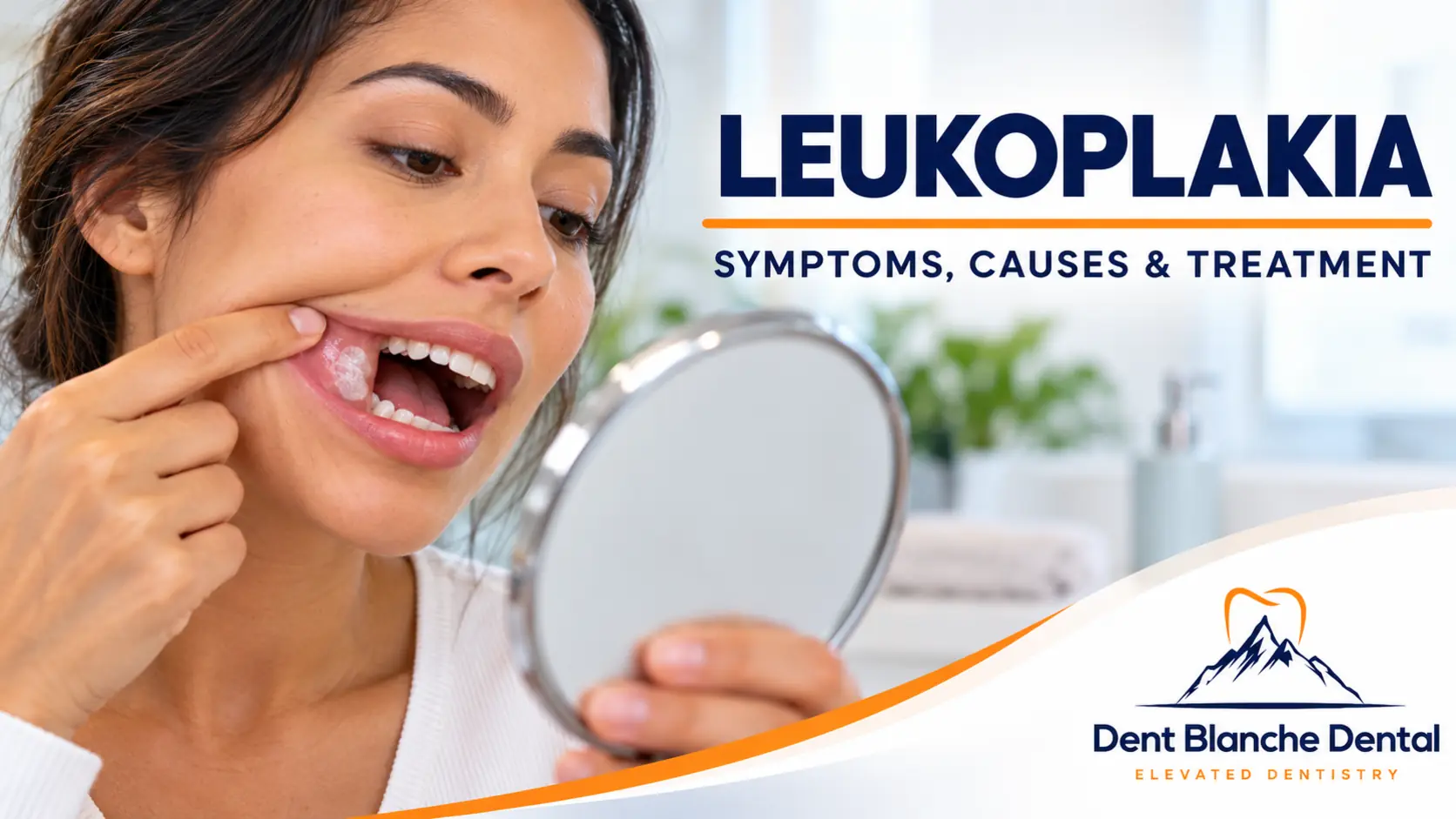
Leukoplakia is a persistent white patch inside the mouth that cannot be scraped off and cannot be explained by another identifiable condition. It may appear on the tongue, gums, cheeks, or floor of the mouth. Although many cases are not cancerous, some contain precancerous changes or may develop into oral cancer. Therefore, any unexplained white patch that persists for more than two weeks should be evaluated by a dentist or oral medicine specialist.
Leukoplakia often causes no pain, making early recognition especially important.
At Dent Blanche Dental in Princeton, NJ, Dr. Radwa Saad provides comprehensive oral examinations and evaluates suspicious oral tissue changes to help patients receive appropriate care.
Overview: What Is Leukoplakia?
Leukoplakia describes a white patch or plaque affecting the oral mucosa that cannot be classified as another recognized oral condition after appropriate evaluation.
Unlike removable debris or certain fungal infections, leukoplakia typically remains attached to the underlying tissue.
It can develop on:
- The sides or underside of the tongue
- The inside of the cheeks
- The gums
- The floor of the mouth
- Other oral surfaces
Not every white patch is leukoplakia. Conditions such as oral candidiasis, frictional keratosis, and oral lichen planus may have a similar appearance.
A professional examination is essential for an accurate diagnosis.
Key Takeaways
- Leukoplakia causes persistent white or grayish patches in the mouth.
- The patches usually cannot be wiped or scraped away.
- Leukoplakia may affect the tongue, cheeks, gums, or floor of the mouth.
- Tobacco exposure is an important risk factor, although cases can occur without tobacco use.
- Most patches are not cancerous, but some show dysplasia or malignant changes.
- Red-and-white patches and certain nonuniform lesions deserve particular attention.
- A biopsy may be necessary to establish a diagnosis and assess risk.
- Long-term monitoring remains important, even after treatment.
What Is Leukoplakia?

Oral leukoplakia is a clinical diagnosis generally made after other causes of persistent white patches have been excluded.
The affected tissue may become thicker because of increased keratin production.
However, appearance alone cannot reliably determine whether a patch is harmless, precancerous, or cancerous.
Some leukoplakias contain epithelial dysplasia, meaning the cells show abnormal changes under a microscope.
This is why dentists take persistent unexplained oral lesions seriously.
Leukoplakia Symptoms
Leukoplakia symptoms vary depending on the size, location, and appearance of the lesion.
Common findings include:
- Persistent white or gray patches
- Thickened or slightly raised tissue
- Rough, wrinkled, or leathery surfaces
- White patches that do not rub off
- Changes in texture or appearance
- Occasional sensitivity to spicy or acidic foods
Many patients experience no discomfort.
Oral Leukoplakia Symptoms That Need Prompt Evaluation
Contact your dentist if you notice:
- A white patch lasting more than two weeks
- A lesion that becomes larger or changes appearance
- Red areas mixed with white tissue
- An ulcer that does not heal
- Unexplained bleeding
- A persistent lump or thickened area
- Difficulty swallowing or moving the tongue
These findings do not necessarily mean cancer, but they require professional assessment.
What Does Leukoplakia Look Like?
Leukoplakia usually appears as a white or gray patch that remains firmly attached to the oral tissue.
Its appearance may be:
- Flat and smooth
- Thick and rough
- Wrinkled or folded
- Slightly raised
- Mixed red and white
Dentists sometimes describe lesions as homogeneous or nonhomogeneous.
Homogeneous leukoplakia generally has a relatively uniform white appearance.
Nonhomogeneous leukoplakia may have irregular, nodular, verrucous, or mixed red-and-white areas.
Certain nonhomogeneous lesions may carry a greater risk of dysplasia or malignant transformation.
Leukoplakia on Gums
Leukoplakia on the gums may appear as a persistent white plaque near the teeth or along the gingival tissue.
However, white gum patches can also result from other conditions.
A dentist must distinguish leukoplakia from frictional changes, inflammatory conditions, and other oral lesions.
White Patches on the Side of the Tongue
White patches on the side of the tongue deserve careful evaluation, particularly if they persist or cannot be wiped away.
The lateral and ventral tongue are important sites during oral cancer examinations.
However, a white tongue patch does not automatically indicate leukoplakia or cancer.
Hairy Leukoplakia
Oral hairy leukoplakia is a distinct condition associated with Epstein-Barr virus.
It commonly produces white, corrugated, or folded patches along the sides of the tongue.
It occurs more often in people with weakened immune systems, although it can appear in other circumstances.
Unlike conventional oral leukoplakia, oral hairy leukoplakia is generally not considered a potentially malignant oral disorder.
However, it may indicate an underlying immune-related concern that requires medical evaluation.
A dentist or physician can help distinguish it from other causes of white tongue lesions.
Leukoplakia Causes
The exact cause of oral leukoplakia is not always known.
However, several factors are associated with its development.
Tobacco Use
Smoking and smokeless tobacco are important risk factors.
Long-term exposure to tobacco products can contribute to changes in the oral mucosa.
Stopping tobacco use may allow some lesions to improve, although others persist.
Alcohol Consumption
Heavy alcohol consumption is associated with oral cancer risk, especially when combined with tobacco.
However, alcohol exposure does not explain every case of leukoplakia.
Chronic Irritation
Sharp teeth, broken restorations, or poorly fitting dentures can cause white frictional lesions.
Importantly, these lesions are not automatically classified as true leukoplakia.
Your dentist may correct the irritation and reassess the tissue to determine whether further investigation is needed.
Other Factors
Leukoplakia can occur in people who have never smoked.
Age, lesion characteristics, and other clinical factors may influence risk.
Because the cause is not always obvious, persistent white patches should not be self-diagnosed.
Leukoplakia Treatment Options
Leukoplakia treatment depends on the diagnosis, lesion characteristics, biopsy findings, and individual risk factors.
1. Comprehensive Oral Examination
Your dentist examines the affected tissue and reviews your medical and dental history.
The examination may include:
- Location and size of the patch
- Surface texture and color
- Duration of symptoms
- Tobacco and alcohol exposure
- Nearby dental irritation
- Other oral tissue changes
2. Biopsy When Indicated
A biopsy may be recommended to examine the tissue under a microscope.
It can help identify:
- Benign tissue changes
- Epithelial dysplasia
- Other oral diseases
- Oral cancer
A biopsy is especially important when a lesion is suspicious, persistent, or changing.
3. Address Contributing Factors
Your dental team may recommend:
- Stopping tobacco use
- Reducing alcohol exposure
- Correcting irritating dental restorations
- Adjusting poorly fitting dentures
- Maintaining good oral hygiene
Removing an irritant does not eliminate the need to investigate a suspicious lesion.
4. Removal or Specialist Treatment
Some lesions may require surgical excision or another specialist-directed approach.
Treatment decisions depend on biopsy findings, lesion location, size, and overall risk.
Even after removal, leukoplakia can recur, and oral cancer risk may persist.
5. Long-Term Monitoring
Regular follow-up examinations are important.
Your dentist or oral medicine specialist may recommend periodic reassessment and repeat biopsy if the lesion changes.
There is no single treatment that guarantees prevention of future oral cancer.
Leukoplakia vs. Other White Patches in the Mouth
Several oral conditions can resemble leukoplakia.
| Condition | Typical appearance | Important distinction |
|---|---|---|
| Leukoplakia | Persistent white patch that does not scrape off | May require biopsy and monitoring |
| Oral thrush | Creamy white plaques, often removable | Usually related to fungal infection |
| Canker sore | Painful ulcer with a pale center and red border | Usually heals within one to two weeks |
| Oral lichen planus | White lace-like lines or patches | Inflammatory condition requiring evaluation |
| Frictional keratosis | White thickening near a source of irritation | May improve after the irritation is removed |
| Oral hairy leukoplakia | Corrugated white patches on the tongue’s sides | Associated with Epstein-Barr virus |
Because these conditions overlap in appearance, a clinical examination is more reliable than online photographs.
When Should You See a Dentist for White Patches in Your Mouth?
Schedule an oral examination if a white patch:
- Persists longer than two weeks
- Cannot be wiped away
- Increases in size
- Develops red areas
- Becomes ulcerated or bleeds
- Appears with a lump or persistent discomfort
Do not attempt to scrape, burn, or remove a suspicious patch yourself.
Early evaluation can help identify the condition and determine whether a biopsy or referral is needed.
Leukoplakia FAQs
What Is Leukoplakia?
Leukoplakia is a persistent white patch in the mouth that cannot be explained by another recognized condition after evaluation. Some lesions may contain precancerous cellular changes.
What Are the Symptoms of Leukoplakia?
Common leukoplakia symptoms include white or gray patches, thickened tissue, and a rough surface. Many lesions are painless.
What Causes Leukoplakia?
Tobacco is an important risk factor. However, leukoplakia can also occur in people without known tobacco exposure, and the exact cause may remain unclear.
Is Leukoplakia Cancerous?
Leukoplakia is not automatically cancer. However, some lesions contain dysplasia or can develop into oral cancer. A biopsy may be needed to assess the tissue.
Is Leukoplakia Contagious?
No. Conventional oral leukoplakia is not contagious and cannot be transmitted through kissing or sharing utensils.
Does Leukoplakia Go Away on Its Own?
Some white lesions may improve after an irritant is removed or tobacco use stops. However, persistent true leukoplakia requires professional monitoring, even if symptoms are absent.
Is Leukoplakia Painful?
Usually not. Some patients experience mild irritation or sensitivity, but many notice only a visible patch.
Leukoplakia vs. Thrush: What’s the Difference?
Thrush often produces removable creamy plaques, while leukoplakia typically cannot be scraped away. However, certain fungal lesions may also remain attached, so examination is necessary.
Leukoplakia vs. Canker Sore: What’s the Difference?
A canker sore is usually a painful ulcer that heals within a couple of weeks. Leukoplakia is generally a persistent white plaque rather than an ulcer.
Is Leukoplakia an Autoimmune Disease?
No. Leukoplakia itself is not classified as an autoimmune disease. Other conditions, including oral lichen planus, may resemble it.
What Can I Do for Leukoplakia Self-Care?
Avoid tobacco, limit alcohol, maintain good oral hygiene, and attend recommended examinations. Do not try to remove persistent white patches at home.
What Are the Available Leukoplakia Treatments?
Treatment may include eliminating risk factors, biopsy, specialist assessment, surgical removal when indicated, and long-term monitoring.
The Bottom Line
Leukoplakia is a persistent white patch in the mouth that should not be ignored.
Although many lesions are not cancerous, some contain abnormal cells or may develop into oral cancer.
A professional oral examination—and a biopsy when indicated—helps determine the diagnosis and appropriate treatment.
If you notice a white patch that lasts longer than two weeks, schedule a dental evaluation.
Concerned About White Patches in Your Mouth? Visit Dent Blanche Dental
At Dent Blanche Dental in Princeton, NJ, Dr. Radwa Saad and our team provide comprehensive oral examinations and evaluate unusual changes in oral tissues.
If you notice persistent white patches on your tongue, gums, or cheeks, we can assess the area and recommend appropriate follow-up or specialist referral.
Early evaluation is an important step in protecting your oral health.
Schedule Your Dental Appointment
Call: 609-890-1888
Dent Blanche Dental
3640 Lawrenceville Road
Princeton, NJ 08540